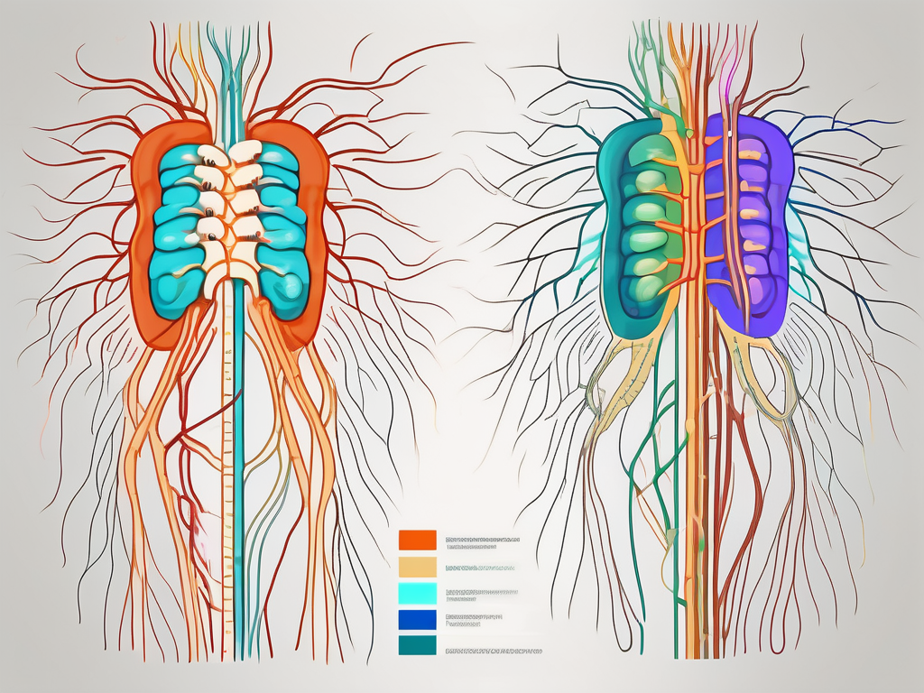
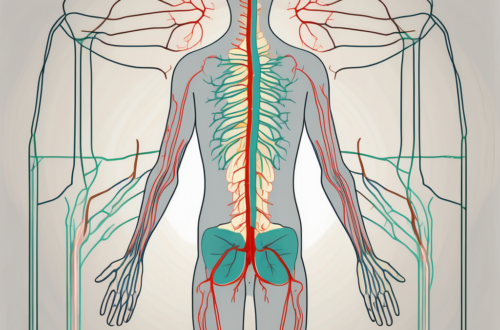
The nervous system is an intricate network that plays a crucial role in controlling and coordinating various bodily functions. Understanding its complexities is essential in grasping the mechanisms underlying our body’s responses to different stimuli. One particular aspect that has intrigued scientists for years is the exit points of presynaptic sympathetic or parasympathetic nerve fibers from the spinal cord. These nerve fibers are essential in transmitting signals from the central nervous system (CNS) to different parts of the body, influencing vital physiological processes.
Understanding the Nervous System
Before diving into the intricacies of nerve fiber exit points, it is essential to have a basic understanding of the nervous system. The nervous system comprises two main components: the CNS and the peripheral nervous system (PNS). The CNS is composed of the brain and spinal cord, while the PNS includes all the nerves running throughout the body.
Communication within the nervous system occurs through specialized cells called neurons. Neurons transmit electrical signals, also known as action potentials, which allow for the transfer of information from one part of the body to another. These signals are crucial in coordinating various bodily functions and maintaining homeostasis.
Neurons are fascinating cells that come in different shapes and sizes. Some neurons have long extensions called axons, which can reach incredible lengths, allowing them to transmit signals over long distances. Other neurons have short, branching extensions called dendrites, which receive signals from neighboring neurons. These intricate connections between neurons form complex networks that enable the nervous system to process and respond to information.
The Role of the Spinal Cord in the Nervous System
Now, let’s focus on the spinal cord, a vital component of the CNS. The spinal cord acts as a link between the brain and the rest of the body, serving as a conduit for nerve fibers. It plays a crucial role in transmitting sensory information from the body to the brain and relaying motor signals from the brain to the muscles.
Imagine a highway system where cars are constantly traveling back and forth, transporting goods and people. In a similar way, the spinal cord serves as a busy highway for nerve signals, ensuring that information flows smoothly between the brain and the body. Without the spinal cord, our ability to sense the world around us and control our movements would be severely impaired.
Additionally, the spinal cord is responsible for integrating reflex actions that occur without conscious thought. When you touch a hot surface and instinctively withdraw your hand, it is the spinal cord that orchestrates this protective response, bypassing the need for the brain’s involvement.
Reflexes are fascinating examples of the spinal cord’s efficiency. They are rapid, involuntary responses that help protect our bodies from harm. For instance, when you step on a sharp object, your leg automatically jerks away before you even consciously register the pain. This reflex action, mediated by the spinal cord, allows for swift and protective responses, preventing further injury.
Sympathetic vs. Parasympathetic Nerve Fibers: A Brief Overview
Within the peripheral nervous system, two branches of the autonomic nervous system, namely the sympathetic and parasympathetic divisions, have distinct roles in regulating bodily functions.
The sympathetic nervous system is responsible for activating the body’s “fight or flight” response. It increases heart rate, dilates blood vessels, and mobilizes energy reserves to prepare the body for action in times of stress or danger.
Imagine you are walking alone in the woods, and suddenly you hear a loud rustling in the bushes. Your heart starts pounding, your breathing quickens, and your muscles tense up. These physiological changes are orchestrated by the sympathetic nervous system, preparing you to either confront the threat or flee from it.
On the other hand, the parasympathetic nervous system promotes the “rest and digest” state. It slows down heart rate, enhances digestion, and conserves energy. The parasympathetic division is active during times of relaxation and promotes the body’s recuperation.
Think about a lazy Sunday afternoon when you are lying on the couch, reading a book, and enjoying a warm cup of tea. Your heart rate slows down, your breathing becomes calm, and your body enters a state of relaxation. These restorative processes are facilitated by the parasympathetic nervous system, allowing your body to recover and recharge.
The sympathetic and parasympathetic divisions of the autonomic nervous system work in harmony, balancing each other’s effects to maintain optimal bodily functions. This delicate interplay ensures that our bodies can respond appropriately to different situations, whether it’s facing a threat or finding moments of rest and rejuvenation.
Anatomy of the Spinal Cord
Now that we have a fundamental understanding of the nervous system and its components, let’s explore the anatomical structure of the spinal cord.
The spinal cord is a long, cylindrical bundle of nerves that extends from the base of the brain, down the vertebral column, until the lumbar spine. It is protected by the bony vertebral column, providing structural support and shielding it from potential injury.
The spinal cord is divided into different regions, each responsible for specific functions. These regions include the cervical, thoracic, lumbar, and sacral segments. Each region gives rise to nerve fibers that control specific areas of the body.
The cervical region, located in the neck area, contains nerve fibers that innervate the upper limbs, including the arms, hands, and fingers. These fibers allow us to perform intricate movements and feel sensations in our upper extremities.
The thoracic region, situated in the chest area, is responsible for controlling the muscles and receiving sensory information from the trunk and abdominal region. It plays a crucial role in maintaining posture and regulating vital functions such as breathing and heart rate.
The lumbar region, found in the lower back, is associated with nerve fibers that control the lower limbs, including the legs and feet. These fibers enable us to walk, run, and perform various movements with our lower extremities.
The sacral region, located at the base of the spine, is responsible for innervating the pelvic organs, bladder, and reproductive organs. It also plays a role in controlling bowel movements and sexual function.
The Path of Nerve Fibers in the Spinal Cord
Within the spinal cord, nerve fibers follow specific pathways, allowing for efficient communication between the CNS and the peripheral tissues. Two main types of nerve fibers exist in the spinal cord: ascending and descending fibers.
Ascending fibers carry sensory information from the body to the brain, allowing us to perceive touch, temperature, pain, and other sensations. These fibers relay important messages, informing the brain of various stimuli and facilitating appropriate responses.
For example, when you touch a hot stove, the sensory receptors in your skin send signals through the ascending fibers to the brain. The brain then interprets this information as pain and quickly sends a message back down the spinal cord to withdraw your hand from the stove, protecting you from further harm.
Descending fibers, on the other hand, transmit motor signals from the brain to the muscles, coordinating voluntary movements. They play a vital role in executing the commands initiated by the brain.
Imagine you want to pick up a glass of water. Your brain sends a message through the descending fibers, instructing the muscles in your arm and hand to contract and coordinate the necessary movements. Without these descending fibers, you would not be able to perform voluntary actions.
In addition to their primary functions, the spinal cord and its nerve fibers also contribute to reflex actions. Reflexes are rapid, involuntary responses to specific stimuli that do not require conscious thought or involvement of the brain.
For example, when you accidentally touch a hot surface, your hand automatically pulls away before you even realize what happened. This reflex action is coordinated by the spinal cord, which quickly processes the sensory information and sends a motor response to protect your body.
In conclusion, the spinal cord is a complex and vital part of the nervous system. Its anatomical structure and the pathways of its nerve fibers allow for efficient communication between the brain and the rest of the body. Understanding the anatomy of the spinal cord is crucial in comprehending how it functions and how it contributes to our everyday movements and sensations.
Exit Points of Presynaptic Nerve Fibers
Now that we have established the fundamentals of the nervous system and the spinal cord, let’s delve into the exit points of presynaptic sympathetic or parasympathetic nerve fibers. These exit points are crucial in determining how these nerve fibers communicate with the body’s various organs and tissues.
Exit Points of Sympathetic Nerve Fibers
Sympathetic nerve fibers exit the spinal cord through distinct pathways. Axons from sympathetic neurons originate from the thoracic and lumbar regions of the spinal cord, forming a chain of interconnected ganglia called the sympathetic chain. From these ganglia, sympathetic nerve fibers extend towards different organs and tissues, allowing for targeted messaging and regulation of physiological responses.
As sympathetic nerve fibers exit the spinal cord, they form a complex network that extends throughout the body. These fibers travel along the spinal nerves, branching off at various points to reach their target destinations. The sympathetic chain, which runs parallel to the spinal cord, plays a vital role in coordinating the activities of these nerve fibers.
One significant exit point of sympathetic nerve fibers is the superior cervical ganglion. Situated at the base of the skull, this ganglion receives input from the sympathetic chain and sends out fibers to innervate structures in the head and neck. These structures include the dilator muscles of the iris, which control the size of the pupil, and the sweat glands of the face and scalp.
Another important exit point is the celiac ganglion, located in the abdomen. From here, sympathetic fibers extend to various abdominal organs, including the liver, stomach, and intestines. These fibers play a crucial role in regulating blood flow, digestive processes, and other physiological functions within the abdominal cavity.
Exit Points of Parasympathetic Nerve Fibers
In contrast, parasympathetic nerve fibers exhibit a unique exit pattern from the spinal cord. Unlike the sympathetic division, the parasympathetic division originates both in the brainstem and the sacral region of the spinal cord.
In the cranial region, fibers from the parasympathetic division exit through cranial nerves, such as the vagus nerve, which innervates organs in the head, neck, and thoracic cavity. The vagus nerve, also known as the wandering nerve, is the longest cranial nerve and has branches that extend to various organs, including the heart, lungs, and digestive system.
Within the sacral region, parasympathetic fibers emerge from the sacral spinal cord and extend to the pelvic organs. These fibers exit through the sacral nerves and innervate structures such as the bladder, reproductive organs, and the lower gastrointestinal tract. The parasympathetic division plays a crucial role in promoting rest and relaxation, as well as regulating various bodily functions in the pelvic region.
It is important to note that while sympathetic and parasympathetic nerve fibers have distinct exit points, they often work in conjunction to maintain homeostasis and ensure the proper functioning of the body. The balance between these two divisions is essential for overall health and well-being.
The Role of Presynaptic Nerve Fibers in the Body
The exit points of presynaptic nerve fibers hold significant implications for the body’s overall functioning. These nerve fibers are responsible for transmitting signals from one neuron to another, allowing for communication and coordination within the nervous system. Let’s explore the functions of sympathetic and parasympathetic nerve fibers in more detail.
Functions of Sympathetic Nerve Fibers
Sympathetic nerve fibers play a crucial role in preparing the body for stressful situations. When faced with a threat or danger, the sympathetic nervous system is activated, triggering a cascade of physiological responses, collectively known as the “fight or flight” response.
During this response, various adaptations occur in the body to ensure survival. One of the key changes is an increase in heart rate, which allows for a greater supply of oxygenated blood to be pumped to the muscles. This increased blood flow helps to enhance physical performance and reaction time, enabling the individual to either fight off the threat or flee from it.
In addition to an increased heart rate, the sympathetic nervous system also causes elevated blood pressure. This rise in blood pressure helps to ensure that an adequate amount of oxygen and nutrients are delivered to the vital organs and tissues, further supporting the body’s response to the stressful situation.
Furthermore, sympathetic nerve fibers cause the dilation of the airways, allowing for increased airflow to the lungs. This enhanced respiratory function ensures that the body receives a sufficient amount of oxygen, which is essential for maintaining optimal performance during times of stress.
Another important effect of sympathetic activation is the enhanced release of glucose from energy stores. This increase in blood sugar levels provides the body with a readily available source of fuel, allowing for sustained energy production during the fight or flight response.
In summary, sympathetic nerve fibers play a vital role in preparing the body for stressful situations. The physiological responses triggered by the sympathetic nervous system help to optimize physical performance, ensuring the individual’s survival in threatening or dangerous circumstances.
Functions of Parasympathetic Nerve Fibers
On the other hand, parasympathetic nerve fibers promote a state of relaxation and restoration. When the body is no longer under stress, the parasympathetic division of the autonomic nervous system is activated, resulting in various physiological responses that counterbalance the effects of sympathetic activation.
One of the primary functions of the parasympathetic nervous system is to decrease heart rate. By slowing down the heart rate, the body conserves energy and allows for a state of rest and recovery. This decrease in heart rate also helps to lower blood pressure, reducing the strain on the cardiovascular system.
In addition to its effects on heart rate, the parasympathetic division stimulates digestion. It promotes the secretion of digestive enzymes and increases the motility of the gastrointestinal tract, facilitating the breakdown and absorption of nutrients from food. This enhanced digestive function ensures that the body receives the necessary nutrients for growth, repair, and overall well-being.
Furthermore, parasympathetic activation enhances the elimination of waste products from the body. It stimulates the contraction of smooth muscles in the urinary and digestive systems, promoting the excretion of waste materials and maintaining homeostasis.
Overall, the functions of parasympathetic nerve fibers are essential for the body’s restorative processes. By promoting relaxation, digestion, and waste elimination, the parasympathetic division helps to conserve energy, repair tissues, and maintain optimal health and well-being.
Implications for Neurological Health and Disorders
The exit points of presynaptic nerve fibers from the spinal cord are essential for maintaining proper neurological function. Disruption or dysfunction of these exit points can lead to various neurological disorders and have significant implications for overall health.
How Nerve Fiber Exit Points Affect Neurological Function
Altered exit points of nerve fibers can disrupt the correct transmission of signals to the organs and tissues they innervate. This can result in uncoordinated responses, leading to a range of symptoms and potential complications.
For example, a misalignment of parasympathetic nerve fibers innervating the pelvic organs may lead to issues with urinary or reproductive function. Similarly, disruption of sympathetic nerve fibers could affect the body’s response to stress, potentially resulting in disorders such as hypertension or cardiovascular dysfunction.
Disorders Related to Nerve Fiber Exit Points
Several neurological disorders may arise from abnormalities at the exit points of presynaptic nerve fibers. Examples include autonomic dysreflexia, a condition characterized by an exaggerated sympathetic response in individuals with spinal cord injuries, and dysautonomia, a dysfunction of the autonomic nervous system that affects multiple bodily functions.
It is important to note that diagnosing and managing disorders related to nerve fiber exit points requires the expertise of healthcare professionals. If you experience any symptoms or concerns related to your nervous system, it is crucial to seek medical advice and consult with a qualified healthcare provider.
Conclusion
Understanding the exit points of presynaptic sympathetic or parasympathetic nerve fibers from the spinal cord provides valuable insights into the functioning of the nervous system. These exit points allow for targeted communication with organs and tissues, ensuring proper physiological responses and maintaining homeostasis.
The intricate interplay between the sympathetic and parasympathetic divisions is vital in coordinating the body’s responses to various internal and external stimuli. Disruption of these exit points can lead to a wide range of neurological disorders, emphasizing the importance of proper diagnosis and management.
As with any medical or neurological concern, always consult with a healthcare professional for personalized advice and guidance. By working together with healthcare providers, we can ensure optimal neurological health and well-being.


