The innervation of parasympathetic nerve fibers in the heart is a complex process that plays a crucial role in maintaining normal cardiac function. Understanding the intricate workings of the parasympathetic nervous system is essential for comprehending the physiological processes that occur within the heart.
Understanding the Parasympathetic Nervous System
The parasympathetic nervous system is one of the two main divisions of the autonomic nervous system, responsible for regulating a variety of bodily functions, including heart rate, digestion, and urination. Unlike the sympathetic nervous system, which is responsible for the “fight or flight” response, the parasympathetic nervous system predominantly acts to restore and conserve energy.
The parasympathetic nervous system originates from the brainstem and sacral region of the spinal cord. It consists of two main cranial nerves – the vagus nerves, which play a vital role in heart regulation, among other functions.
The parasympathetic nervous system is a fascinating and intricate network that extends throughout the body, ensuring the smooth functioning of various organs. Let’s delve deeper into the role of the parasympathetic nervous system in heart function and explore the anatomy of this remarkable system.
Role of the Parasympathetic Nervous System in Heart Function
Within the heart, the parasympathetic nervous system exerts control over the rate and force of contraction, as well as other critical aspects of cardiac function. By releasing acetylcholine, the key neurotransmitter involved in parasympathetic signaling, parasympathetic nerve fibers slow down the heart rate and decrease cardiac output.
This regulatory mechanism helps maintain the balance between the sympathetic and parasympathetic branches of the autonomic nervous system. Without this delicate balance, the heart’s rhythm and efficiency could be compromised, leading to various cardiovascular disorders.
It is fascinating to note that the parasympathetic nervous system’s influence on heart function extends beyond regulating heart rate. It also affects the electrical activity of the heart, ensuring the coordination of atrial and ventricular contractions.
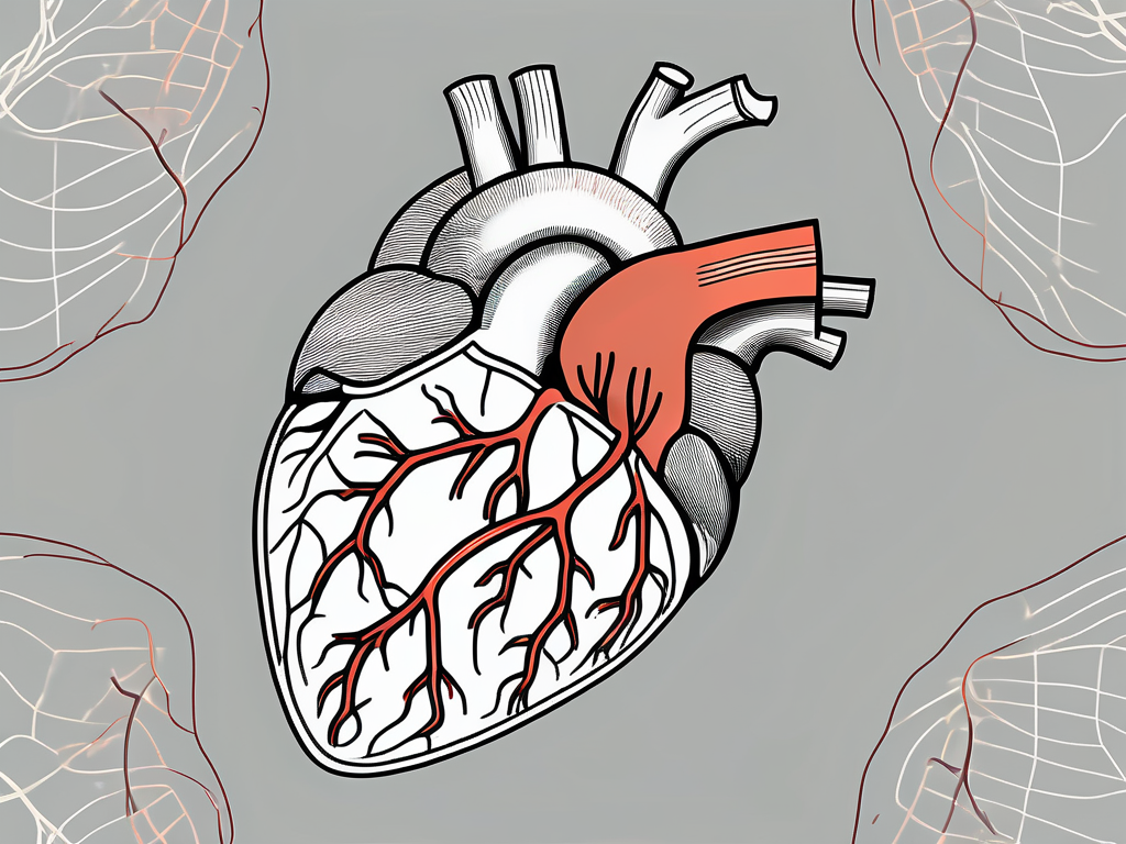
The parasympathetic nerve fibers primarily innervate the sinoatrial (SA) and atrioventricular (AV) nodes, which are responsible for initiating and coordinating the heart’s rhythmic contractions. The presence of parasympathetic nerve fibers in these regions allows for precise control over heart rate and the coordination of atrial and ventricular contractions.
Moreover, the parasympathetic nervous system’s impact on heart function is not limited to the physical aspects. It also plays a role in emotional regulation, as it is responsible for the calming and relaxing response that helps reduce stress and anxiety.
Anatomy of the Parasympathetic Nervous System
The parasympathetic nervous system consists of a network of nerve fibers that extend from the brainstem and sacral region to various organs throughout the body, including the heart. In the heart, parasympathetic nerve fibers primarily innervate the sinoatrial (SA) and atrioventricular (AV) nodes, which are responsible for initiating and coordinating the heart’s rhythmic contractions.
These nerve fibers form neurotransmitter-releasing junctions with specialized cells known as pacemaker cells, which generate electrical signals that regulate the heart’s electrical activity. The presence of parasympathetic nerve fibers in these regions allows for precise control over heart rate and the coordination of atrial and ventricular contractions.
Understanding the intricate anatomy of the parasympathetic nervous system helps us appreciate the complexity and precision with which it regulates heart function. The network of nerve fibers extends throughout the body, ensuring that every organ receives the necessary parasympathetic input for optimal functioning.
It is remarkable to think about how this intricate system evolved over millions of years, fine-tuning the balance between the sympathetic and parasympathetic branches of the autonomic nervous system to maintain homeostasis and promote overall well-being.
Parasympathetic Nerve Fibers and Heart Innervation
Heart innervation by parasympathetic nerve fibers involves a complex series of interactions and signal transduction processes. The primary mechanisms through which parasympathetic control is exerted in the heart include the process of heart innervation and the impact of parasympathetic nerve fibers on heart rate.
Process of Heart Innervation
The process of heart innervation begins with the release of acetylcholine from parasympathetic nerve terminals onto specialized receptor cells located in the SA and AV nodes. These cells, commonly referred to as cholinergic cells, possess specific receptors, such as muscarinic receptors, that bind acetylcholine and initiate downstream signaling events.
Upon binding, muscarinic receptors activate intracellular signaling cascades, leading to a reduction in the rate of electrical impulses generated by the SA node. As a result, heart rate decreases, allowing for proper coordination of atrial and ventricular contractions. This intricate regulation ensures optimal cardiac output and overall cardiovascular health.
Furthermore, the process of heart innervation is not limited to the SA and AV nodes. Parasympathetic nerve fibers also extend to the atria and ventricles, forming a dense network that allows for precise control over the electrical activity of the heart. This network ensures that the atria and ventricles contract in a synchronized manner, maximizing the efficiency of blood pumping.
In addition to their role in heart rate regulation, parasympathetic nerve fibers also play a crucial role in modulating the strength of cardiac contractions. Through the release of acetylcholine, these fibers can inhibit the activity of the sympathetic nervous system, which is responsible for increasing heart rate and contractility. This delicate balance between sympathetic and parasympathetic inputs ensures that the heart functions optimally under different physiological conditions.
Impact of Parasympathetic Nerve Fibers on Heart Rate
Parasympathetic nerve fibers have a profound impact on heart rate modulation. Through the release of acetylcholine and subsequent activation of muscarinic receptors, parasympathetic stimulation slows down the heart rate, contributing to what is commonly referred to as “vagal tone.”
In situations where the parasympathetic nervous system is overactive or dominant, such as during periods of rest and relaxation, heart rate can drop to below-normal levels, a condition known as bradycardia. While bradycardia can be asymptomatic in some individuals, it may cause symptoms such as fatigue, dizziness, and shortness of breath. If experiencing symptoms, it is important to consult a healthcare professional for proper evaluation and guidance.
It is worth noting that parasympathetic nerve fibers do not solely influence heart rate. They also have an impact on other aspects of cardiac function, such as the conduction of electrical impulses through the AV node. By slowing down the conduction velocity, parasympathetic stimulation allows for proper coordination between the atria and ventricles, ensuring efficient blood flow and preventing arrhythmias.
Moreover, parasympathetic nerve fibers can also affect the contractility of the heart muscle. Through the release of acetylcholine, these fibers can decrease the force of contraction, which can be beneficial in certain situations, such as reducing the workload on the heart during periods of rest. However, excessive parasympathetic stimulation can lead to decreased contractility, resulting in reduced cardiac output and compromised cardiovascular function.
In summary, the role of parasympathetic nerve fibers in heart innervation is crucial for maintaining proper cardiac function. Through the release of acetylcholine and activation of muscarinic receptors, these fibers regulate heart rate, conduction velocity, and contractility. The delicate balance between sympathetic and parasympathetic inputs ensures optimal cardiovascular performance under various physiological conditions. Understanding the intricate mechanisms of parasympathetic control in the heart provides valuable insights into the complex nature of cardiac regulation.
Mechanisms of Parasympathetic Control in the Heart
The mechanisms underlying parasympathetic control in the heart are complex and involve the interplay of various neurotransmitters, receptors, and signal transduction pathways. Understanding these mechanisms is crucial for uncovering potential therapeutic targets and improving the treatment and prevention of heart diseases.
Parasympathetic control in the heart is a finely tuned process that involves the coordinated release and interaction of multiple neurotransmitters. Acetylcholine, as mentioned earlier, is the primary neurotransmitter involved in parasympathetic signaling within the heart. It is released by parasympathetic nerve fibers and binds to muscarinic receptors, initiating a cascade of intracellular events that ultimately influence heart rate and contraction strength.
But acetylcholine is not the only neurotransmitter involved in parasympathetic control. Nitric oxide, another important player, also participates in regulating heart activity. Nitric oxide functions as a vasodilator, helping to relax blood vessels, reduce cardiac workload, and enhance overall cardiovascular function. Its release and interaction with other neurotransmitters contribute to the precise regulation of heart activity.
Neurotransmitters Involved in Parasympathetic Control
Acetylcholine, as mentioned earlier, is the primary neurotransmitter involved in parasympathetic signaling within the heart. It binds to muscarinic receptors, initiating a cascade of intracellular events that ultimately influence heart rate and contraction strength.
In addition to acetylcholine, other neurotransmitters, such as nitric oxide, also participate in parasympathetic control. Nitric oxide functions as a vasodilator, helping to relax blood vessels, reduce cardiac workload, and enhance overall cardiovascular function. The coordinated release and interaction of these neurotransmitters contribute to the precise regulation of heart activity.
Furthermore, recent research has uncovered the involvement of other neurotransmitters, such as vasoactive intestinal peptide (VIP) and adenosine, in parasympathetic control of the heart. VIP acts as a potent vasodilator and plays a role in regulating heart rate and contractility. Adenosine, on the other hand, helps to regulate coronary blood flow and myocardial oxygen consumption.
Receptors and Signal Transduction in the Heart
The heart contains various receptor subtypes that mediate the effects of parasympathetic nerve fibers. Muscarinic receptors, located on the cholinergic cells in the SA and AV nodes, are particularly critical in transducing the parasympathetic signals.
Upon binding of acetylcholine, these receptors activate intracellular signaling pathways, such as the G-protein coupled receptor (GPCR) pathway, leading to the suppression of cyclic adenosine monophosphate (cAMP) production and subsequent modulation of various ion channels. These ion channels play a vital role in regulating the electrical conduction system of the heart and, ultimately, heart rate.
In addition to muscarinic receptors, other receptor subtypes, such as adenosine receptors and VIP receptors, are also involved in parasympathetic control of the heart. Adenosine receptors, when activated by adenosine, modulate the activity of ion channels and contribute to the regulation of heart rate and contractility. VIP receptors, when stimulated by VIP, initiate a signaling cascade that leads to vasodilation and influences heart rate.
The signal transduction pathways involved in parasympathetic control of the heart are complex and interconnected. In addition to the GPCR pathway, other pathways, such as the phospholipase C (PLC) pathway and the nitric oxide-cyclic guanosine monophosphate (NO-cGMP) pathway, also play important roles. These pathways mediate the intracellular events that ultimately result in changes in heart rate, contractility, and vascular tone.
Overall, the mechanisms of parasympathetic control in the heart are multifaceted and involve the interplay of various neurotransmitters, receptors, and signal transduction pathways. Further research in this field is necessary to fully understand these mechanisms and their potential therapeutic implications for heart diseases.
Clinical Significance of Parasympathetic Innervation in the Heart
Understanding the clinical significance of parasympathetic innervation in the heart is crucial for diagnosing and managing various cardiovascular disorders. Disruptions in parasympathetic control can lead to a wide range of cardiac abnormalities that can significantly impact a person’s health and quality of life.
The parasympathetic nervous system plays a vital role in regulating heart rate, blood pressure, and cardiac function. It acts as a counterbalance to the sympathetic nervous system, which is responsible for the “fight or flight” response. The parasympathetic innervation, also known as the vagus nerve, releases acetylcholine, a neurotransmitter that slows down the heart rate and promotes relaxation.
Parasympathetic Nervous System Disorders and the Heart
In certain conditions, such as autonomic dysfunction or certain types of heart disease, the parasympathetic nervous system may become impaired, leading to an imbalance between sympathetic and parasympathetic influences on the heart.
For example, conditions like autonomic neuropathy, which affects the peripheral nerves, can disrupt parasympathetic signaling and result in heart rate irregularities, exercise intolerance, and blood pressure dysregulation. Similarly, conditions such as atrial fibrillation, where the heart’s electrical signals become disorganized, can be influenced by parasympathetic dysfunction.
It is crucial for individuals experiencing these symptoms or concerns to consult a healthcare professional promptly. Only a qualified medical practitioner can accurately diagnose and guide appropriate treatment strategies.
Diagnosing parasympathetic dysfunction often involves a combination of medical history evaluation, physical examination, and specialized tests such as heart rate variability analysis or autonomic function tests. Treatment approaches may include lifestyle modifications, medications, or interventions targeting the underlying cause of the parasympathetic imbalance.
Therapeutic Approaches Targeting Parasympathetic Innervation
Given the significant impact of parasympathetic innervation on cardiac function, researchers and clinicians are exploring various therapeutic approaches that target this intricate network. These approaches aim to potentiate or restore parasympathetic control in individuals with cardiovascular disorders.
Emerging strategies include targeting specific receptors or enzymes involved in parasympathetic signaling, as well as electrical neuromodulation techniques that directly influence parasympathetic nerve activity. These advancements hold promise for improving heart health and potentially reducing the risk of cardiac complications.
One such approach is vagus nerve stimulation (VNS), which involves the implantation of a device that delivers electrical impulses to the vagus nerve. VNS has shown promising results in managing certain cardiac conditions, such as heart failure and atrial fibrillation. By stimulating the parasympathetic nervous system, VNS can help regulate heart rate, reduce inflammation, and improve overall cardiac function.
Another area of research focuses on developing pharmacological agents that selectively target parasympathetic receptors, such as muscarinic receptors. These agents aim to enhance parasympathetic signaling without affecting other physiological processes. By fine-tuning the parasympathetic control, these medications may offer new treatment options for individuals with cardiovascular disorders.
Furthermore, non-invasive techniques like transcutaneous vagus nerve stimulation (tVNS) are being explored as potential adjunct therapies. tVNS involves the application of electrical stimulation to the skin overlying the vagus nerve, stimulating its activity and promoting parasympathetic function. Preliminary studies have shown promising results in improving heart rate variability and reducing inflammation in individuals with heart disease.
While these therapeutic approaches show great potential, further research is needed to fully understand their effectiveness, safety, and long-term outcomes. Collaborations between researchers, clinicians, and industry partners are essential to advance the field and bring these innovative treatments to clinical practice.
Future Directions in Parasympathetic Heart Innervation Research
The field of parasympathetic heart innervation research is continually evolving, with ongoing studies exploring novel techniques and potential implications for heart disease treatment and prevention.
Emerging Techniques in Studying Heart Innervation
Advancements in imaging technologies and molecular biology techniques have opened new avenues for studying the intricate details of parasympathetic heart innervation. Researchers can now visualize and track individual nerve fibers and specific molecules involved in parasympathetic signaling, enabling a more comprehensive understanding of this complex system.
Additionally, the use of genetically modified animal models and advanced molecular techniques, such as gene editing, helps researchers gain insights into the specific genes and proteins involved in parasympathetic control. These cutting-edge techniques contribute to the identification of potential therapeutic targets and the development of precise interventions.
Potential Implications for Heart Disease Treatment and Prevention
As our knowledge of parasympathetic heart innervation continues to expand, there is growing optimism regarding its potential implications for heart disease treatment and prevention.
By elucidating the mechanisms underlying parasympathetic control, researchers aim to develop targeted therapies that can enhance parasympathetic signaling in individuals with cardiovascular disorders, leading to improved heart function and overall cardiovascular health.
Furthermore, a deeper understanding of parasympathetic heart innervation may provide valuable insights into potential preventive strategies, offering opportunities to intervene at early stages and mitigate the risk of developing heart disease.
Conclusion
The innervation of parasympathetic nerve fibers in the heart is a fascinating area of study that has significant implications for cardiac function and overall cardiovascular health. Understanding the intricate mechanisms that govern parasympathetic control is vital for diagnosing, managing, and potentially treating various heart conditions.
As research progresses and our understanding of parasympathetic innervation improves, we are provided with new opportunities to optimize heart health. It is crucial to continue supporting research efforts in this field, as they hold the potential to revolutionize cardiovascular medicine and improve outcomes for individuals with heart disease.