The human body is a marvel of interconnected systems and networks, where each intricate detail plays a vital role in maintaining our overall health and well-being. One such connection that has fascinated scientists and medical professionals alike is the link between the esophagus and the parasympathetic nerve. In this article, we will delve into the depths of this intricate connection, unraveling the role of the trigeminal nerve in this process.
Understanding the Esophagus and the Parasympathetic Nerve
Before we delve into the specifics of this connection, let’s first gain a comprehensive understanding of the esophagus and the parasympathetic nerve. The esophagus is a muscular tube that connects the throat (pharynx) to the stomach, facilitating the passage of food and liquids. On the other hand, the parasympathetic nerve is a branch of the autonomic nervous system, responsible for regulating many of our internal organs’ functions.
Anatomy of the Esophagus
The esophagus is a hollow, muscular tube measuring approximately 25 centimeters in length. It runs behind the trachea (windpipe) and in front of the spinal column, providing a direct pathway for the passage of food and liquids from the mouth to the stomach. The wall of the esophagus comprises multiple layers, including mucosa, submucosa, muscularis propria, and adventitia or serosa, each playing a unique role in its function.
The innermost layer of the esophagus is the mucosa, which is composed of epithelial cells. This layer secretes mucus to protect the esophageal lining from the abrasive effects of food and liquids. The submucosa lies beneath the mucosa and contains blood vessels, nerves, and glands. It provides support and nourishment to the other layers of the esophagus.
The muscularis propria is the thickest layer of the esophageal wall and is responsible for the peristaltic contractions that propel food towards the stomach. This layer consists of two types of muscle fibers: circular muscles that constrict the esophagus and longitudinal muscles that shorten it. The coordinated contraction and relaxation of these muscles create wave-like movements, pushing food downwards.
Finally, the outermost layer of the esophagus is either adventitia or serosa, depending on its location in the body. The adventitia is a connective tissue layer that attaches the esophagus to surrounding structures, while the serosa is a smooth, slippery membrane that covers the esophagus in the abdominal cavity.
Role of the Parasympathetic Nerve
The parasympathetic nerve is responsible for maintaining a balanced state in our body, often referred to as rest and digest mode. It regulates various important bodily functions, including digestion, heart rate, and breathing. In the context of the esophagus, the parasympathetic nerve plays a crucial role in controlling the muscle contractions (peristalsis) that propel food down towards the stomach.
When we eat, the parasympathetic nerve stimulates the release of acetylcholine, a neurotransmitter that activates the muscles in the esophagus. This activation leads to coordinated contractions of the circular and longitudinal muscles, creating the peristaltic waves necessary for the movement of food. Without the parasympathetic nerve’s influence, the esophagus would not be able to efficiently transport food from the mouth to the stomach.
In addition to its role in peristalsis, the parasympathetic nerve also regulates the secretion of saliva and other digestive enzymes in the esophagus. These enzymes help break down food particles, making them easier to swallow and digest. The parasympathetic nerve ensures that the esophagus is adequately prepared for the arrival of food, facilitating the smooth and efficient digestion process.
Furthermore, the parasympathetic nerve also plays a role in controlling the lower esophageal sphincter (LES), a ring of muscle located at the junction between the esophagus and the stomach. The LES acts as a valve, preventing stomach acid and partially digested food from flowing back into the esophagus. The parasympathetic nerve helps maintain the tone and function of the LES, ensuring that it remains closed when it should be, preventing the occurrence of acid reflux and heartburn.
In conclusion, the esophagus and the parasympathetic nerve work in harmony to facilitate the smooth passage of food from the mouth to the stomach. The intricate anatomy of the esophagus allows for efficient peristalsis, while the parasympathetic nerve controls and coordinates the muscle contractions necessary for this process. Understanding this connection is crucial for comprehending the complex mechanisms behind digestion and maintaining a healthy gastrointestinal system.
The Trigeminal Nerve: A Key Player
Now that we have a basic understanding of the esophagus and the parasympathetic nerve, let’s explore the role of the trigeminal nerve in this intricate connection. The trigeminal nerve, also known as the fifth cranial nerve, is one of the twelve cranial nerves responsible for transmitting sensory information from the face to the brain.
The Trigeminal Nerve Explained
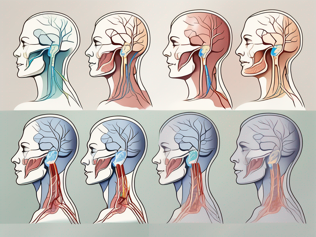
The trigeminal nerve is composed of three branches: the ophthalmic, maxillary, and mandibular branches. It supplies sensation to various areas of the face, including the forehead, cheek, and jaw. Additionally, it plays a crucial role in our ability to chew, speak, and swallow – functions directly related to the esophagus.
Let’s delve deeper into the three branches of the trigeminal nerve. The ophthalmic branch is responsible for transmitting sensory information from the forehead, scalp, and upper eyelid. It allows us to feel sensations such as touch, temperature, and pain in these areas. The maxillary branch, on the other hand, supplies sensation to the lower eyelid, cheek, and upper lip. It enables us to experience various sensations, including pressure, temperature, and pain in these regions. Lastly, the mandibular branch provides sensory information from the lower lip, chin, and jaw. It allows us to feel sensations such as touch, temperature, and pain in these areas.
Its Role in the Esophageal-Parasympathetic Connection
Recent research has shed light on the intricate connection between the trigeminal nerve and the parasympathetic nerve, particularly in relation to the esophagus. It appears that the trigeminal nerve plays a pivotal role in transmitting sensory information between the esophagus and the parasympathetic nerve, influencing the regulation of muscle contractions and overall esophageal function.
Let’s explore how the trigeminal nerve contributes to the esophageal-parasympathetic connection in more detail. When we eat, the trigeminal nerve is responsible for transmitting sensory information from the oral cavity to the brain. This information includes the taste, temperature, and texture of the food we consume. The brain then processes this information and sends signals to the parasympathetic nerve, which in turn regulates the muscle contractions of the esophagus, allowing the food to move smoothly from the mouth to the stomach.
Moreover, the trigeminal nerve also plays a role in the reflexes involved in swallowing. When we swallow, the trigeminal nerve detects the movement of the tongue and the pressure exerted by the food bolus against the oral cavity. It relays this information to the brain, which triggers the appropriate reflexes to facilitate the swallowing process. These reflexes involve the coordinated contraction of various muscles, including those in the esophagus, to propel the food towards the stomach.
Furthermore, the trigeminal nerve is involved in the sensation of heartburn, a common symptom of gastroesophageal reflux disease (GERD). When stomach acid flows back into the esophagus, it can cause a burning sensation in the chest and throat. The trigeminal nerve detects this sensation and transmits it to the brain, allowing us to perceive and respond to the discomfort.
In conclusion, the trigeminal nerve is a key player in the esophageal-parasympathetic connection. Its branches supply sensation to various areas of the face, and it plays a crucial role in chewing, speaking, and swallowing. Additionally, it is involved in transmitting sensory information between the esophagus and the parasympathetic nerve, influencing muscle contractions and overall esophageal function. Understanding the role of the trigeminal nerve in this intricate connection enhances our knowledge of the complex mechanisms involved in digestion and swallowing.
The Esophagus-Parasympathetic Nerve Connection
Now that we understand the individual components of this complex connection, let’s explore how they work together to ensure proper esophageal function and regulation of muscle contractions.
The esophagus, a muscular tube that connects the throat to the stomach, plays a crucial role in the digestion process. It serves as a pathway for food and liquids to travel from the mouth to the stomach, where further breakdown and absorption occur. To ensure the smooth movement of these substances, the esophagus relies on a sophisticated network of nerves, including the parasympathetic nerve.
How the Connection Works
The esophagus and the parasympathetic nerve communicate through a network of nerve fibers and signaling molecules. When food or liquids enter the esophagus, sensory receptors within the esophageal wall send signals through the trigeminal nerve to the brain. These receptors detect the presence of food and initiate a cascade of events to ensure proper digestion.
Upon receiving the signals from the sensory receptors, the brain relays this information to the parasympathetic nerve. The parasympathetic nerve, also known as the “rest and digest” system, is responsible for regulating various bodily functions, including digestion. It coordinates the appropriate muscle contractions needed for the smooth movement of food down the esophagus.
Through the release of neurotransmitters, such as acetylcholine, the parasympathetic nerve stimulates the muscles in the esophageal wall to contract in a coordinated manner. This peristaltic movement propels the food forward, ensuring that it reaches the stomach efficiently. The parasympathetic nerve also relaxes the lower esophageal sphincter, a muscular ring that separates the esophagus from the stomach, allowing food to pass through.
Implications of the Connection
The esophagus-parasympathetic nerve connection has far-reaching implications for our overall health. Any disruption in this connection can lead to a range of gastrointestinal symptoms, including difficulty swallowing, heartburn, and even more severe conditions such as gastroesophageal reflux disease (GERD).
Gastroesophageal reflux disease occurs when the lower esophageal sphincter fails to close properly, allowing stomach acid to flow back into the esophagus. This can cause irritation and inflammation, leading to symptoms such as heartburn, regurgitation, and chest pain. By understanding the intricate connection between the esophagus and the parasympathetic nerve, researchers and healthcare professionals can develop targeted interventions and potential treatment strategies for individuals affected by these conditions.
Furthermore, studying this connection can shed light on the underlying mechanisms of other esophageal disorders, such as achalasia, a condition characterized by the inability of the esophagus to move food into the stomach. By unraveling the complexities of the esophagus-parasympathetic nerve connection, researchers can uncover new insights into the pathophysiology of these disorders and explore innovative therapeutic approaches.
In conclusion, the esophagus-parasympathetic nerve connection is a vital component of our digestive system. Its intricate coordination ensures the proper functioning of the esophagus and the smooth movement of food from the mouth to the stomach. Understanding this connection not only provides valuable insights into gastrointestinal disorders but also opens doors to potential interventions and treatment strategies for individuals suffering from these conditions.
Unraveling the Trigeminal Link
As we delve deeper into the intricacies of the esophagus, parasympathetic nerve, and trigeminal nerve connection, let’s explore the finer details of the trigeminal link and its impact on this complex network.
The trigeminal link, often referred to as the trigeminal nerve, is the largest cranial nerve in the human body. It is responsible for transmitting sensory information from the face, including touch, pain, and temperature, to the brain. However, recent scientific discoveries have revealed that the trigeminal link is not only restricted to sensory transmission but also influences the release of neurotransmitters and neuropeptides within the esophageal wall.
This newfound understanding suggests that the trigeminal nerve not only transmits sensory information but also actively facilitates the communication between the esophagus and the parasympathetic nerve through chemical signaling. This intricate connection plays a crucial role in maintaining the proper functioning of the esophagus and the regulation of various physiological processes.
The Trigeminal Link in Detail
Scientists have conducted extensive research to unravel the complexities of the trigeminal link. Through meticulous studies, they have discovered that the trigeminal nerve fibers extend into the esophageal wall, forming an intricate network that interacts with the parasympathetic nerve fibers.
These nerve fibers are responsible for the transmission of sensory information from the esophagus to the brain, allowing us to perceive sensations such as pain, pressure, and temperature. Additionally, the trigeminal link plays a crucial role in modulating the release of neurotransmitters and neuropeptides within the esophageal wall.
Neurotransmitters, such as acetylcholine, play a vital role in the regulation of esophageal peristalsis, the coordinated muscular contractions that propel food and liquids through the esophagus. The trigeminal link influences the release of acetylcholine, thereby modulating the strength and frequency of esophageal contractions.
Furthermore, neuropeptides, such as substance P and calcitonin gene-related peptide (CGRP), are involved in the regulation of pain and inflammation. The trigeminal link has been shown to influence the release of these neuropeptides within the esophageal wall, potentially playing a role in the perception of esophageal pain and the development of inflammatory conditions.
The Impact of the Trigeminal Link on the Esophagus-Parasympathetic Nerve Connection
Further research is needed to fully unravel the impact of the trigeminal link on the esophagus-parasympathetic nerve connection. However, preliminary studies have indicated that disruptions in the trigeminal link may contribute to the development or exacerbation of esophageal disorders such as achalasia, a condition characterized by impaired esophageal peristalsis.
Achalasia is a rare disorder that affects the ability of the esophagus to move food and liquids into the stomach. It is believed that abnormalities in the trigeminal link may disrupt the proper coordination of esophageal contractions, leading to the symptoms associated with achalasia, such as difficulty swallowing, regurgitation, and chest pain.
Understanding the role of the trigeminal link in the esophagus-parasympathetic nerve connection could potentially lead to new therapeutic approaches for the treatment of esophageal disorders. By targeting the trigeminal link, researchers may be able to develop interventions that restore the proper functioning of the esophagus and alleviate the symptoms associated with these conditions.
In conclusion, the trigeminal link is a fascinating aspect of the esophagus-parasympathetic nerve connection. Its involvement in sensory transmission, as well as the modulation of neurotransmitters and neuropeptides, highlights its significance in maintaining the proper functioning of the esophagus. Further research is warranted to fully comprehend the intricacies of this connection and its potential implications for the diagnosis and treatment of esophageal disorders.
Potential Medical Implications
The intricate connection between the esophagus, parasympathetic nerve, and trigeminal nerve holds immense potential for medical implications, allowing us to better understand and potentially treat a range of gastrointestinal and neurological disorders.
Implications for Gastrointestinal Disorders
Gastrointestinal disorders, such as GERD, achalasia, and dysphagia, can significantly impact an individual’s quality of life. By unraveling the esophagus-parasympathetic nerve connection and the role of the trigeminal nerve within this network, medical professionals can develop more targeted treatment strategies, aiming to alleviate symptoms and improve overall patient outcomes.
Implications for Neurological Disorders
Neurological disorders, including migraines and trigeminal neuralgia, often exhibit comorbidities with gastrointestinal symptoms. Understanding the intricate connection between these two systems can help identify potential underlying mechanisms and facilitate the development of multidisciplinary treatment approaches.
Future Research Directions
While significant progress has been made in unraveling the connection between the esophagus, parasympathetic nerve, and trigeminal nerve, there is still much to explore. Here are a few potential areas of study that could expand our knowledge and deepen our understanding of this intricate network.
Unanswered Questions
Despite the progress made in recent years, researchers continue to grapple with several unanswered questions. For instance, the exact mechanisms through which the trigeminal link affects esophageal function remain to be fully elucidated. Further investigation is also required to determine whether targeting the trigeminal nerve could be a potential treatment modality for esophageal disorders.
Potential Areas of Study
Exploring the influence of other cranial nerves and neural networks on the esophagus-parasympathetic nerve connection could also shed light on this intricate network. Additionally, studying the development and plasticity of this connection throughout life may provide insights into potential therapeutic interventions that could aid individuals experiencing disrupted esophageal function.
While our understanding of the connection between the esophagus and the parasympathetic nerve, specifically via the trigeminal link, has come a long way, the complexity of this connection reminds us of the vastness that still lies ahead in the field of medical research. As we continue to unravel these intricacies, it is essential to consult with medical professionals and seek their expertise to fully comprehend and address any health concerns or symptoms one may experience. Together, we can expand our knowledge and chart new paths towards improved healthcare outcomes.


