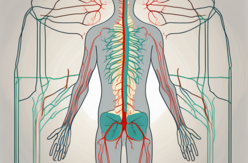
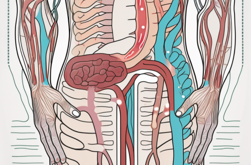
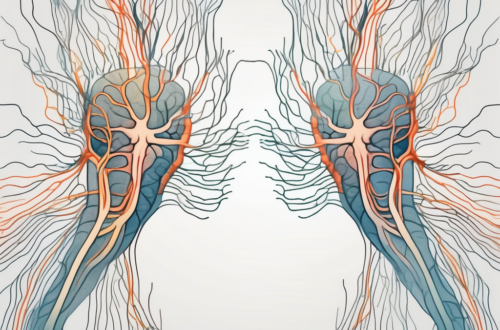
The location of preganglionic parasympathetic nerve cell bodies is a critical aspect of understanding the autonomic nervous system and its functioning within the human body. To comprehend this aspect fully, it is essential to delve into the intricacies of the autonomic nervous system and the role of the parasympathetic nervous system within it.
Understanding the Autonomic Nervous System
The autonomic nervous system is a division of the peripheral nervous system that plays a vital role in regulating various involuntary bodily functions. It controls processes such as heart rate, digestion, breathing, and glandular secretions. The autonomic nervous system is further divided into two branches: the sympathetic nervous system and the parasympathetic nervous system. Both branches work together to maintain balance and homeostasis within the body.
The autonomic nervous system is a complex network of nerves that extends throughout the body, connecting various organs and tissues. It is responsible for coordinating and controlling the body’s automatic responses, allowing us to adapt to different situations and environments.
The sympathetic nervous system is often referred to as the “fight or flight” response system. It is responsible for preparing the body for action in times of stress or danger. When activated, it increases heart rate, dilates blood vessels, and releases stress hormones such as adrenaline. These physiological changes help us respond quickly and effectively to potential threats.
The Role of the Parasympathetic Nervous System
The parasympathetic nervous system primarily acts to restore the body to a resting state after periods of stress or activity. It promotes relaxation, conserves energy, and enhances digestion and elimination. This branch of the autonomic nervous system is responsible for stimulating bodily functions during rest and digest situations.
When the parasympathetic nervous system is activated, it slows down heart rate, constricts blood vessels, and increases digestive activity. This allows the body to conserve energy and focus on processes such as digestion, absorption of nutrients, and elimination of waste products.
The parasympathetic nervous system is active during periods of relaxation and sleep. It helps us unwind, rejuvenate, and restore our energy levels. Without the parasympathetic nervous system, our bodies would constantly be in a state of heightened alertness, leading to exhaustion and potential health problems.
The Structure of the Autonomic Nervous System
The autonomic nervous system is organized in a hierarchical manner, with nerve cell bodies located in specific regions. The preganglionic neurons of both the sympathetic and parasympathetic branches originate within the central nervous system.
From the central nervous system, these preganglionic neurons extend out to ganglia, which are clusters of nerve cell bodies located outside the central nervous system. The sympathetic ganglia are located close to the spinal cord, while the parasympathetic ganglia are located near or within the target organs.
Upon reaching the ganglia, the preganglionic neurons synapse with postganglionic neurons. These postganglionic neurons then extend to the target organs, where they release neurotransmitters that activate or inhibit specific functions.
The structure of the autonomic nervous system allows for efficient and coordinated communication between the central nervous system and the target organs. It ensures that the appropriate responses are initiated in a timely manner, allowing the body to adapt and maintain optimal functioning.
The Anatomy of Preganglionic Parasympathetic Nerve Cells
The preganglionic parasympathetic nerve cells play a fundamental role in transmitting signals from the central nervous system to the effector organs of the body. They are located in specific regions and project their axons to connect with ganglia, which are clusters of nerve cell bodies outside the central nervous system.
These specialized nerve cells, known as preganglionic parasympathetic neurons, are an integral part of the autonomic nervous system. They form a crucial link between the central nervous system and the peripheral organs, ensuring the smooth regulation of bodily functions.
Within the central nervous system, these neurons are found in distinct regions, including the brainstem and the sacral spinal cord. Each region serves a specific purpose in coordinating the parasympathetic response in different parts of the body.
The Function of Preganglionic Neurons
Preganglionic neurons transmit signals from the central nervous system to the postganglionic neurons located in various organs and tissues throughout the body. These neurons act as intermediaries, relaying messages and enabling communication between different parts of the body.
When a stimulus is detected by the central nervous system, such as a change in environmental temperature or the need for digestion, preganglionic neurons are activated. They then propagate the signal to the postganglionic neurons, which carry out the necessary response in the target organs.
For example, in the case of digestion, preganglionic parasympathetic neurons in the brainstem send signals to the postganglionic neurons located in the gastrointestinal tract. These postganglionic neurons, in turn, stimulate the release of digestive enzymes, increase blood flow to the digestive organs, and promote peristalsis, ensuring efficient digestion and nutrient absorption.
The Pathway of Preganglionic Neurons
The pathway of preganglionic neurons starts in the brainstem and sacral spinal cord. In the brainstem, the cranial nerves serve as conduits for the parasympathetic outflow. These cranial nerves, including the oculomotor nerve, facial nerve, glossopharyngeal nerve, and vagus nerve, carry the preganglionic fibers to their respective target organs.
Each cranial nerve has a specific role in regulating different bodily functions. For instance, the oculomotor nerve innervates the muscles responsible for controlling pupil size and lens shape, allowing for proper visual focus. The facial nerve controls salivation, tear production, and taste sensation. The glossopharyngeal nerve is involved in swallowing, salivation, and taste perception. Lastly, the vagus nerve, the longest cranial nerve, provides parasympathetic innervation to the heart, lungs, and gastrointestinal tract, among other organs.
In contrast, the sacral spinal cord provides innervation to pelvic organs and structures. Preganglionic parasympathetic neurons originating from the sacral region control functions such as bladder contraction, bowel movements, and sexual arousal. These neurons travel through the pelvic nerves to reach their target organs, ensuring proper coordination of the parasympathetic response in the pelvic region.
Understanding the pathway of preganglionic neurons is crucial for comprehending the intricate network of communication within the autonomic nervous system. The precise connections and distribution of these neurons allow for the precise regulation of bodily functions, maintaining homeostasis and ensuring optimal physiological responses.
The Location of Preganglionic Parasympathetic Nerve Cell Bodies
Understanding the location of preganglionic parasympathetic nerve cell bodies is essential for comprehending the functioning of the parasympathetic nervous system and its impact on various bodily functions.
The parasympathetic nervous system, often referred to as the “rest and digest” system, plays a crucial role in maintaining homeostasis in the body. It counterbalances the sympathetic nervous system, which is responsible for the “fight or flight” response. The parasympathetic system primarily regulates activities that occur during rest, such as digestion, urination, and sexual arousal.
One of the key aspects of understanding the parasympathetic nervous system is knowing the location of preganglionic parasympathetic nerve cell bodies. These cell bodies are found in specific regions of the body, including the cranial nerves and the sacral spinal cord.
The Cranial Nerves and Their Role
The cranial nerves are an integral part of the parasympathetic outflow system. These nerves emerge directly from the brain and extend to various structures in the head and neck region. Several cranial nerves, including the oculomotor, facial, glossopharyngeal, and vagus nerves, contain preganglionic parasympathetic fibers that innervate specific organs.
The oculomotor nerve, for example, carries preganglionic parasympathetic fibers that control the constriction of the pupil and the accommodation of the lens for near vision. The facial nerve, on the other hand, innervates the salivary glands, controlling the production and secretion of saliva. The glossopharyngeal nerve plays a role in regulating the salivary glands as well, along with the parotid gland, which is responsible for producing saliva.
The vagus nerve, the longest cranial nerve, has extensive parasympathetic innervation throughout the body. It supplies preganglionic fibers to various organs, including the heart, lungs, liver, stomach, and intestines. The vagus nerve is crucial for maintaining proper gastrointestinal function, controlling heart rate, and regulating breathing.
The Sacral Spinal Cord and Its Importance
In addition to the cranial nerves, the sacral spinal cord, located in the lower back region, is also involved in housing preganglionic parasympathetic nerve cell bodies. These cells innervate structures in the pelvis, such as the bladder and genitalia.
The parasympathetic fibers originating from the sacral spinal cord play a vital role in controlling bladder function. They stimulate the contraction of the bladder wall muscles while simultaneously relaxing the muscles of the bladder neck and urethra, allowing for the voluntary release of urine.
Furthermore, the preganglionic parasympathetic fibers from the sacral spinal cord are responsible for the sexual arousal response. They contribute to the dilation of blood vessels in the genitalia, leading to increased blood flow and engorgement of erectile tissues in both males and females.
Overall, understanding the location of preganglionic parasympathetic nerve cell bodies provides valuable insights into the intricate workings of the parasympathetic nervous system. It highlights the diverse range of bodily functions that rely on the parasympathetic system for regulation and control.
The Significance of Preganglionic Parasympathetic Nerve Cell Bodies
The presence and functioning of preganglionic parasympathetic nerve cell bodies have significant implications for bodily functions and overall well-being.
The parasympathetic nervous system, which is responsible for the body’s rest and digest response, relies on the activity of preganglionic parasympathetic nerve cell bodies. These specialized cells are located in specific regions of the central nervous system, including the brainstem and the sacral spinal cord.
The Impact on Bodily Functions
Preganglionic parasympathetic nerve cell bodies play a crucial role in regulating essential bodily functions such as heart rate, digestion, urinary function, sexual arousal, and the secretion of various glands. Their proper functioning is vital for maintaining optimal health and balance within the body.
For example, when these nerve cell bodies are activated, they release neurotransmitters that stimulate the heart to slow down, promoting a state of relaxation and reducing stress. Additionally, they stimulate the release of digestive enzymes and increase blood flow to the digestive organs, facilitating efficient nutrient absorption and digestion.
Furthermore, the parasympathetic nervous system’s influence on urinary function ensures the proper contraction and relaxation of the bladder muscles, allowing for effective urine storage and elimination. In terms of sexual arousal, the parasympathetic nervous system promotes the release of nitric oxide, which leads to the dilation of blood vessels in the genital area, facilitating increased blood flow and engorgement.
The Role in Disease and Disorders
Disruptions or damage to preganglionic parasympathetic nerve cell bodies can lead to various disorders and medical conditions. Dysfunctions in the parasympathetic nervous system can manifest as gastrointestinal disorders, cardiovascular abnormalities, and urogenital dysfunctions.
For instance, damage to the preganglionic parasympathetic nerve cell bodies that innervate the gastrointestinal tract can result in conditions such as gastroparesis, where the stomach muscles fail to properly contract, leading to delayed emptying and digestive disturbances.
In terms of cardiovascular abnormalities, dysfunction in the parasympathetic nervous system can contribute to conditions like bradycardia, where the heart rate becomes abnormally slow. This can lead to symptoms such as fatigue, dizziness, and fainting.
Urogenital dysfunctions associated with preganglionic parasympathetic nerve cell body damage can include urinary incontinence, erectile dysfunction, and difficulties with sexual arousal. These conditions can significantly impact an individual’s quality of life and may require medical intervention to manage symptoms effectively.
If you suspect any abnormalities or experience persistent symptoms related to the parasympathetic nervous system, it is crucial to consult with a medical professional for a proper diagnosis and guidance. They can conduct a thorough evaluation, which may involve various tests and examinations to determine the underlying cause of the issue and develop an appropriate treatment plan.
Future Research Directions in Neuroanatomy
Despite significant advancements in neuroanatomy, there is still much to explore and uncover regarding the location and functioning of preganglionic parasympathetic nerve cell bodies.
The Challenges in Studying Nerve Cell Bodies
Studying nerve cell bodies can be intricate due to their location and microscopic nature. Researchers face challenges in accessing these structures and obtaining comprehensive understanding. However, advancements in imaging techniques, such as MRI and advanced microscopy, continue to provide new insights.
One of the main challenges in studying nerve cell bodies is their intricate network within the central nervous system. These cell bodies are densely packed and interconnected, making it difficult to isolate and study individual cells. Additionally, their microscopic size requires specialized techniques and equipment to visualize and analyze.
Another challenge arises from the delicate nature of nerve cell bodies. These structures are highly sensitive to changes in their environment, making it crucial to handle them with extreme care during experimentation. Any slight disturbance or damage can alter their functioning, leading to inaccurate results.
Furthermore, the location of preganglionic parasympathetic nerve cell bodies adds another layer of complexity. These cell bodies are distributed throughout the body, making it challenging to access and study them in vivo. Researchers often rely on post-mortem studies or animal models to investigate these structures.
The Potential for New Discoveries
Ongoing research and advancements in neuroscience hold the promise of uncovering new discoveries about the location and functioning of preganglionic parasympathetic nerve cell bodies. These discoveries may lead to a deeper understanding of various physiological processes and potential therapeutic interventions.
With the advent of cutting-edge technologies, researchers are now able to delve deeper into the intricacies of nerve cell bodies. Advanced imaging techniques, such as functional MRI and high-resolution microscopy, allow for non-invasive visualization of these structures in living organisms. This opens up new avenues for studying their functioning in real-time and under different physiological conditions.
Moreover, the integration of molecular biology techniques with neuroanatomy research has the potential to revolutionize our understanding of nerve cell bodies. By identifying specific genes and proteins associated with these cells, researchers can unravel the molecular mechanisms underlying their development, connectivity, and functioning.
Additionally, advancements in computational modeling and data analysis are enhancing our ability to interpret complex neuroanatomical data. By combining large-scale datasets and sophisticated algorithms, researchers can uncover patterns and relationships within the intricate network of nerve cell bodies, providing valuable insights into their organization and functioning.
In conclusion, understanding the location of preganglionic parasympathetic nerve cell bodies is crucial for comprehending the functioning of the parasympathetic nervous system and its impact on bodily functions. Further research and exploration in this field hold the potential for new discoveries and advancements in neuroanatomy. If you have any concerns or questions regarding your bodily functions, it is always advisable to consult with a qualified healthcare professional for appropriate guidance and care.


